Tuesday, 2 November 2010

you've been eliminated

unlike reality shows, you can't vote a drug off your system if you want to eliminate it.

a drug gets eliminated by either ZERO ELIMINATION CONSTANT or FIRST ORDER ELIMINATION.

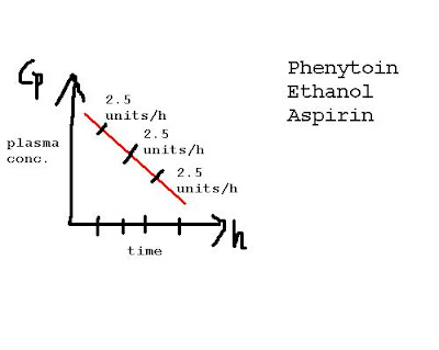
a zero order elimination is when the plasma concentration of a drug decreases linearly with time:


a drug with first order elimination has a rate of elimination proportional to the drug concentration:


from the graph you can see, the more drug in the system (the higher the plasma conc.), the faster the drug is eliminated.

drugs are also metabolized in the system propr to elimination. there are 2 phases of this metabolism.

phase I
includes reduction, oxidation and hydrolysis. it yields water soluble metabolites. sometimes a drug is activated after phase I metabolism. the cytochrome p450 system is connected to phase I. old patients lose phase I first!

phase II @ conjugation includes acetylation, glucuronidation, sulfation and yields inactive metabolites that are renally excreted.

pharma calculations recall

let's go over the 4 most basic pharma equations:

  1. Vd (volume of distribution) = concentration of drug given (IV) / concentration of drug in plasma

    you have to take bioavailability (how much of the drug is subjected to first pass metabolism) into account if drug is not given IV.
  2. clearance = (0.7 x Vd)/ t1/2
                             
                                or    K x Vd  (where K = elimination constant = 0.7 / t1/2 )
  3. loading dose = concentration of steady state x Vd
    *conc of steady state = desired conc
  4. maintenance dose = (concentration of steady state x clearance) / bioavailability                    
so for maintenance doses, you can see from the equation that it is determined by clearance. why do you need to know this? clearance rates are affected in patients with renal/ hepatic disease. in a patient with renal disease, maintenance dose is DECREASED (coz clearance is decreased), but Vd remains the same (does not depend on kidney function), therefore loading dose remains the same.

t1/2 is the amount of time it takes for a drug to reach 1/2 it's plasma concentration.

if you infuse a drug, it takes 4t1/2s to achieve a steady state of 94%. likewise, it take 4t/12s for 94% of said drug to be eliminated.

(remember that 1t1/2 = 50% of conc, 2t1/2s = 75% [50% + 25%], 3t1/2s = 87.5% and 4 t1/2s = 94%)

on my next post, we'll touch elimination constants and drug metabolism.

Saturday, 30 October 2010

more on Tc

what simple test tests for Tc function?anergy panel skin test. examples include: PPD, Candida, Trichophyton, tetanus toxoid, saline control). it is an example of delayed HS (HS 4). for a test to be +ve, Tc function must be preserved. remember, that HS1-3 is mediated by B cells.


what interferes with a anergy panel?
  • corticosteroids (topical/ high dose systemic)
  • anticoagulants (you get a skin induration because fibrin deposition, which is absent in settings of anticoagulants use)
  • technique (i.e doctor's mistake at not injecting the antigen intradermally)
  • infection (HIV = no functioning Tc, EBV, TB, leprosy)
  • autoimmune disease (R.A, SLE, sarcoid)
  • malnutrition
  • pregnancy
  • malignancy (eg: Hodgkin's lymphoma)
what assays test for Tc?
flow cytometry for CD3, 4, 8, 45 and Tc receptor

oh, dear

apparently i've spelt minuscule wrong all along.

i really didn't know that until spell check put underlined it with that wavy red line.

cell markers

CD3: all Tc
CD4: Th c
         60-70% of Tc in blood and lymphoid tissues.
         receptor for HIV
CD8: marker for Tk c (cytotoxic Tc)
CD19, 20: Bc
CD16, 56: NK c

Tc markers have functional significance, whereas Bc markers have a maturational significance.

Thursday, 21 October 2010

immuno revisited

let's go over some factoids regarding ILs (interleukins)...those itty-bitty things that make immuno seem impossible at times!
  • type of IL that boosts T helper cell?
    IL-2
  • btw, normal Tc to Bc ratio?
    3:1
  • causes an increase in temperature?
    TNF-alfa and IL-1
  • revs up IgA?
    IL-5
  • IL involved in helminthic infections?
    IL-5 (it revs up both IgA [intestinal mucosa] and eosinophils = activated at helminthic infections)
  • responsible for endotoxin septic shock and causes cachexia in malignancies?
    TNF-alfa
  • bonus question: where does TNF-alfa come from?
    it is secreted by macrophages
  • another point if you can tell me the mechanism for how it causes cachexia?
    it inhibits lipoprotein lipase in adipose tissues
  • interesting connection you can make:
    TNF-alfa activates IL-2 and Bc
  • major, major must know fact: at HS1 (hypersensitivity type 1), which IL is the main culprit? think before looking at answer!
    did you guess IL5? (boo). TNF-alfa? (booooo).
    the answer is: IL-4, which then revs up IgE --> anaphylactic shock!
  • IL responsible for activating stem cells?
    IL-3 (i remember this with a silly mnemonic: tree of life --> stem c are new life --> IL3)
  • acute phase of inflamation...2 main ILs?
    IL-1 (remember...fever!) and IL-6 (acts as GM-CSF)
  • mature monocytes secrete what CKs?
    first, you must answer...what are mature monocytes? MACROPHAGES! (back to the histo books for you if you didn't know that), so what did i say earlier? mphags secrete TNF-alfa and IL-1
  • where are mphags fixed in tissues and what mediator activates them?
    recall that in liver cells, mphags = Kuppfer cells and C5a is needed to get them a moving again.
it is said the human brain at one time can retain 7 active items in its memory. i've given you 14 for now :) time to give these factoids a good home in your cerebellum :)

icannotbreathe!

ARDS (acute respiratory distress syndrome) carries a 50% death rate. stuff you should know or die:

  • main causes?
    ischemic shock, DIC, endotoxin release (septic shock), illicit drug use, acute pancreatitis, breathing in really hot air
  • main cell responsible?
    NEUTROPHILS!
  • so, one of the features of ARDS is pulmonary edema. how do we differentiate this from say...cardiogenic pulmonary edema?
    pulmonary capillary wedge pressure (LV) *please review the physio part of this if you feel like you've never heard this before*
    it is low in ARDS and high in cardiogenic pulm. ed.

Thursday, 7 October 2010

adrenals gone bad

let's do a review of some endocrine pathologies, shall we?

starting with my most favourite: Cushing's syndrome.

what is it? an increase of cortisol due to various reasons, which can be divided into:
  • exogenous: steroid intake (iatrogenic)
  • endogenous:
    1. cushing's disease (70%). a pituitary adenoma secreting excess ACTH, which in turn increases secretion of cortisol from the adrenals. serum shows an increase in both ACTH and cortisol.
    2. ectopic ACTH, made by non pituitary tissues (small cell lung cancer, bronchial carcinoids). serum will show an increase in both ACTH and cortisol.
    3. adrenal adenoma, carcinoma, nodular hyperplasia. ACTH levels would be LOW.
how do we test for it and differentiate between the three?

Dexamethasone suppression test
 
  • in healthy individuals, cortisol levels drop after a low dose of dexamethasone (the increase in cortisol, inhibits secretion of ACTH, which means there's no stimulation for the adrenals to produce cortisol)
  • in an ACTH producing pituitary tumor (1), the cortisol levels increase after a low dose, but decrease after a high dose.

    now, you have a tissues producing excess ACTH, with almost no negative feedback from cortisol. that's why a low dose of exogenous steroid (dexamethasone) fails to inhibits further ACTH secretion and therefore the levels of cortisol still appear high. when a bigger dose of dexamethasone is administered, the negative feedback system is triggered to decrease the cortisol level.

    so, low dose dexamethasone, cortisol levels high. high dose dexamethasone, cortisol levels drop.

  • ectopic ACTH producing tissues (2) and adrenal adenomas (3) differ in the sense that they are not influenced by the hypothalamus-pituitary-adrenal axis, and therefor not subjected to the negative feedback system.

    so, both a low and high dose of dexamethasone will result in high cortisol levels.
now onto HYPERALDOSTERONISM. two types exist:

  1. primary (Conn's syndrome): aldosterone secreting tumor (uni/bilatera) that results in hypertension, hypoK+, metabolic alkalosis, low plasma renin.

  2. secondary: kidney perceives low intravascular volume (due to renal artery stenosis, CHF, CRF, cirrhosis, nephrotic syndrome) and this results in an overactive renin-angiotensin-aldosteron activation. key point here is: PLASMA RENIN WOULD BE HIGH.
treatment? spironolactone, an aldosterone antagonist (its MOA as a K+ sparring diuretic).

next up: ADDISON'S.

  • chronic adrenal insufficiency due to adrenal hypoplasia (primary = problem at adrenal) . results in total absence of hormones from all three layers of the adrenal cortex (primary deficiency of cortisol and aldosterone = hypotension).

    skin pigmentation occurs due to increase MSH, a by product of ACTH, which is increased because the pituitary tries stimulating the non responding adrenals.

    so increased ACTH, low adrenal hormones (salt, sugar, sex), high MSH = hyperpigmentation (bronze).
  • secondary adrenal insufficiency = problem at pituitary. so ACTH would be LOW, no skin hyperpigmentation and no hyperkalemia.
that's all for today folks. stay tuned =)

Sunday, 19 September 2010

Acute Post Transfusion Hemolysis

So a recently transfused patient presents with Acute Renal Failure, low BP, Pruritus and so on and so forth. An emergency life threatening situation. So how'd you treat? Any emergency situation, list out the patient's pathologies and prioritize them in decreasing order of how life threatening they are to the patient.

The patient's problems are: The post transfusion hemolysis itself, and its complications: Acute Renal Failure and low BP & hypoperfusive shock.

Treat the low BP first : Cardiac inotropes - Dopamine is probably ideal, with minimal renal effects; Rheopolyglucin

IV Fluids

Dialysis - If available : Probably the best option.

IV glucocorticosteroids

A suitable anticoagulant

Furosemide in high doses, to increase GFR*

Plasmapheresis if necessary


Edit: After a thorough dressing down by the mother, I realized that furosemide is contraindicated unless systolic BP > 100. So, furosemide only after BP is stabilized. Apologies for that boo-boo.

Friday, 10 September 2010

bilevel positive pressure ventilation (BiPAP) in CHF patients

diuresis is paramount in patients with CHF that have AFib. anti arrhythmic drugs can help bring down the rate, but would not ineffective minus diuresis (atrial stretch from fluid overload causes the patient to go into Afib).


a trial of BiPaP can go a long way in stabilizing the patient (decreases afterload, increases preload and increases CO) while you are awaiting the effects of diuresis.

did you know?

A hepatic abscess caused by Klebsiella, has an association with DM and septic endopthalmitis.

Sunday, 11 July 2010

deal breaker

insulin causes K+ to shift into the cells thereby decreasing the extracellular K level.

remember:

INsulin = K+ INto cells.

that's why insulin is used in the treatment of hyperkalemia.

there's another vital concept regarding the insulin-K+ relationship which has slipped my mind and i would need to look up.

Monday, 28 June 2010

mental defense mechanisms - psych stuff

first off, sorry for the lack of updates. am up to my ears with catching up on studies. however, my notebook is already half filled with REALLY GOOD stuff i'll upload over my summer break.

for now, here's an easy way to remember all the different type of defense mech, using the husband and wife examples.

the dude that came up with this is a genius!


  1. Husband attracted to another woman goes back and make love to his wife.
  2. Husband attracted to another woman goes back and offer flowers to his wife.
  3. Husband attracted to his student then he becomes her mentor.
  4. Husband having affair with another woman justifies to himself by saying this will make me appreciate my wife more.
  5. Husband attracted to another woman then he thinks that his wife is cheating on him.
  6. Husband attracted to another woman and explains to his friend that how sexual desires are instinctual and cannot be controlled.
  7. Husband attracted to another woman then goes and write about how bad cheating husbands are in the newspaper.
  8. Husband fought with his wife then went fighting at a football game.
  9. Husband told of grave diagnosis and tells his wife "Honey, I need your help without you I cannot fight".
Answer key
  1. Displacement
  2. Undoing
  3. Sublimation
  4. Rationalization
  5. Projection
  6. Intellectualization
  7. Reaction formation
  8. Displacement
  9. Regression
reaction formation = doing the extreme opposite of what you long/ desire for.
undoing = not extreme opposite, somewhat same direction as desire (eg: giving flowers = sign of love and affection).

Wednesday, 23 June 2010

let's play some more

  1. you get a 30 y.o. male who has been experiencing severe pruritus (itching) for the past 2 weeks. he has a history of ulcerative colitis (UC) for the past seven years, and is on sulfasalazine and cortisone enemas. he has diffuse excoriations on his extremities and trunk. his labs reveal a mild iron deficiency anemia and normal electrolytes. LFTs are normal, except for an increase in alkaline phosphatase: 322 U/L (normal <110 U/L). What is the most likely explanation for his symptoms?

    Primary Sclerosing Cholangitis (PSC)
    the patient has had longstanding UC and has now developed pruritus in the setting of an elevated alkaline phosphatase. this sclerosing process involves both the intra- and extrahepatic ducts and is diagnosed by ERCP.

    PSC occurs most often in young men and is commonly associated with IBDs, especially UC. PSC has a triad of progressive fatigue, pruritus and jaundice. there may also be upper quadrant pain, fever, hepatosplenomegaly or cirrhosis.

    complications of PSC include progression to decompensated cirrhosis, PHT, ascites, and liver failure. treatment is generally supportive and include antibacterial treatment for superimposed bacterial cholangitis.

    Primary biliary cirrhosis also presents with pruritus and an elevated alkaline phosphatase, but it is typically seen in middle-aged women and has no association with ulcerative colitis, thus ruling it out from the differential.

  2.  this case is a gem: a 64-year-old man who is currently undergoing chemotherapy, experiences the occasional nausea and vomiting, for which he is given IV prochlorperazine to help ease the symptoms. after several days of therapy, he complains that he feels restless, agitated and he cannot stop moving his legs. what medication should you have given to him at the beginning of his therapy to prevent this reaction? choose between Haloperidol and Lorazepam.

    LORAZEPAM.

    why? because this patient has akathisia, a syndrome characterized by unpleasant sensations of "inner" restlessness that manifests itself with an inability to sit still or remain motionless (definition from wiki). this the feeling of restlessness sometimes occurs as a side effect of neuroleptic drugs, such as prochlorperazine and haloperidol. akathisia can be prevented by administering IV benzodiazepines, such as lorazepam, concomitantly with the neuroleptic drug. this is especially vital in a patient whose immune system and metabolic activity is compromised.

  3. here's an obstretic pearl. let's say a pregnant woman comes for her antenatal check up. everything is a ok, except her urine dipstick shows bacteriuria (asymptomatic because she doesn't have dysuria, increased frequency, a temperature or increased urgency). whaddya do?

    asymptomatic bacteriuria is present in about 5% of pregnant women. because it may cause preterm delivery/low birth weight, it is vital that all pregnant women be screened for asymptomatic bacteriuria early in the pregnancy and be treated if affected by it.

    e. coli is the main organism most of the time. other gram-negative organisms (e.g: klebsiella, enterobacter, and proteus species) and gram-positive cocci (e.g: enterococci and group B strep) may be responsible as well.

    treatment choices include: trimethoprim-sulfamethoxazole, nitrofurantoin, and cephalexin. ampicillin and amoxicillin may be administered as well (bear in mind though that e.coli may be resistant to these drugs).

    10 days after completing the course of antibiotics, the patient should have a follow-up urine culture to make sure the causative agent of bacteriuria has been eradicated.

  4. ok, bonus question. you have to be able to answer this (or it's time to hit those patho books again): pitituary adenomas, what hormone level will be elevated?

    PROLACTIN!

  5. all right, i know most of us didn't like public health/ public service medicine too much as a subject, but statistics play a big role in diagnosing a patient right too.

    let's go back to 2 basic points before i give you the question:


    • sensitivity = (positive)/ (positive + false negative) x100.
      a sensitive screening test detects DISEASED individuals.
      usually used to screen for disease with low prevalance.
      remember: sensitivity rules out. (SnOUT).
    • specificity = (negative)/ (negative +  false positive) x 100.
      a specific screening test detects HEALTHY individuals.
      usually used as a confirmatory test after a positive sensitive test.
      remember: specificity spins in (SpIN).

    now...you get a 70 y.o male come to you to complain about his very red and swollen right toe, that hurts severely. he is not on any medications and denies abusing alcohol. you know it's gout, but which method will you use to make that diagnosis? pick from colchicine response or checking his uric acid levels.

    colchine response. u.a levels may be elevated in gout, but it is also increased at tumor lysis syndrome and other diseases. so the results won't be specific enough.

    so give oral colchicine hourly until the patient develops improvement in joint pain and inflammation.


    of course the most specific method of diagnosis, would be joint aspiration and seeing negatively birefringent needle shaped crystals under the microscope. but think CHEAP, get the right diagnosis and your chief of medicine will love you for saving his the bucks.


    until the next round folks =)

let's play the diagnosis game


1.            you have a 50-year-old man with ringing in his ears and he feels the room spinning around him. he also tells you his hearing in his left ear has slowly gotten worse. he says that this all began a while ago, along with a slight feeling of unsteadiness. he has no chronic medical conditions and does not take any medications. examination shows nystagmus. what is the most likely diagnosis?

Dx: meniere's disease, which is characterized by tinnitus, vertigo, and progressive hearing loss (his hearing got worse slowly). it is thought to be related to a degeneration of the vestibular and cochlear hair cells. treatment includes bed rest, a low-salt diet, dimenhydrinate, cyclizine or meclizine. 
perhaps you considered it being benign positional vertigo, but recall that, BPV is characterized by paroxysmal vertigo and nystagmus. it is brought on by certain changes in position and hearing loss is not present. therefor, you can rule it out of the differential.

2.            an 18-year-old male student is admitted for psychosis. he is put in seclusion because he is highly aggressive and a threat to others. he was given haloperidol intramuscularly twice so far. suddenly, he has acute torticollis and twitching of the mouth and face on that side. what reaction did this patient most likely have?

acute dystonia
, an involuntary spasm of a particular group of muscles that can involve the neck, jaw, tongue, eyes, or the entire body. it can be an early adverse effect of antipsychotics, and it is more common in younger men. it is more common with typical antipsychotics. the treatment of choice is parenteral administration of anticholinergics.

 3.            a 15-year-old, obese boy complains of persistent knee pain for several weeks and came into your office, limping. he sits in the examining table with the sole of the foot on the affected side pointing to the other leg. however, physical examination is normal for the knee, but shows limited hip motion. when the hip is flexed, the leg goes into external rotation and cannot be rotated internally. which of the following is the most likely diagnosis: avascular necrosis of the femoral head OR slipped capital femoral epiphysis?

slipped capital femoral epiphysis, an orthopedic emergency. it has a classic clinical picture: a chubby male, early teens, who is limping and cannot rotate his leg internally. also, this hip pathology will produce knee pain, but the knee is normal on physical examination.

avascular necrosis is seen in younger children, around 6 years of age.

Friday, 18 June 2010

dermatomes!

  • C6-C8: 
    • C6 covers the thumb and index finger (medial half). to remember this, make the number 6 with your left hand by touching your index finger to your thumb.
    • then, remember C8 the is the lateral digit (baby finger) and half of the 4th
    • which leaves C7 getting whats inbetween C6 and C8
  • L5 vs S1
    • intervetebral discs herniation happens at this level
    • L5 affects the big toe
      • L5 = Largest of the 5
      • L5 root is compressed by a herniated disc at the L4-L5 level
    • S1 affects the smallest toe
      • S1 = the smallest one
      • S1 root is compressed at the L5-S1 level
    • L1 = Skin overlying the inguinal ligament
      • L for ligament, 1 for 1nguinal
    • S5 = Anus
      • S5 is the doodie hive (got this off the net: doodie being another word for poop and hive = house...so erm yeah)
  • Others:
    • T10 = Umbilicus
      • Bellybut-TEN
    • C3, 4, 5 keeps the diaphragm alive
    • L4 = Knee
      • L4 hits the floor (when kneeling)

Thursday, 17 June 2010

M & M & M & M

what?    
    • Mycoplasma pneumoniae (causes atypical pneumonia aka walking pneumonia: insidious onset, headache, non productive cough, diffuse infiltrate, respiratory symptoms don't dominate)
who? 
    • Military and Minors and Misfits (prisoners)
how? 
    • IgM (which causes cold agglutination and lysis of rbc which can lead to hemolytic anemia)
    • bullous Myringitis (bullae on the tympanic membrane)
Tx = erythromycin or tetracycline. resistant to penicillin because? come on gander a guess (think MOA of penicillin and you'd understand immediately).
if you guessed coz it has no cell wall, good for you =) it's the only bacterial membrane with cholestrol. so it can't be seen on gram stain.

Wednesday, 16 June 2010

vitamin B

to a med student, knowing their B1, 2, 3, 6, 12 is as intrinsic as a preschooler knowing their A, B, Cs. sometimes the difference between an A grade and a B grade, is getting the right vitamin B type.


B1 thiamine
In thiamine pyrophosphate (TPP) a cofactor for:

  • Pyruvate DH (glycolysis)
  • Alpha ketoglutarate DH (TCA)
  • Transketolase (HMP)
  • Branched chained aa DH
     
At malnutrition or alcoholism (secondary malnutrition and malabsorption)

Impaired glucose breakdown leading to ATP depletion, so first organs affected are the brain and heart (highly aerobic tissues).

Wernicke-Korsakoff syndrome.
Wernicke:
  • confusion
  • opthalmoplegia,
  • ataxia
Korsakoff:
  • memory loss
  • confabulation
  • personality change
Beriberi:
§         wet beriberi (high output HF, dilated cardiomyopathy, edema)
§         dry beriberi (polyneuritis, symmetrical muscle atrophy)

mnemonic: ber1ber1
B2 riboflavin
Cofactor in oxidation and reduction (FADH2)
2 Cs:
  • Cheilosis
  • Corneal vascularization

mnemonic:
riboFlavin = FAD, FMN= 2 ATP
B3 niacin
  • Redox rxn (NAD+, NADP+)
  • Derived from tryptophan
  • Synthesis requires B6


*excess: facial flushing (seen at pharmacological doses of treating hyperlipidemia)
3 Ds of pellagra:
  • Dermatitis
  • Diarrhea
  • Dementia
Severe deficiency caused by:
1.      Hartnup’s disease (decreased tryptophan absorption)
2.      Malignant carcinoid syndrome (increased tryptophan metabolism)
3.      INH (decreased B6)

B5 pantothenate
Cofactor for acyl transfers and FA synthase
  • Dermatitis
  • Enteritis
  • Alopecia
  • Adrenal insufficiency
B6 pyridoxine
·        Required for synthesis of B3 from tryptophan
·        Converted to pyridoxal phosphate, a cofactor for:
«     transamination (eg: ALT, AST)
«     DC reactions
«     Glycogen phosphorylase
«     Cystathione synthesis
«     Heme synthesis


§         Convulsions
§         Hyperirritability
§         Peripheral neuropathy
§         Sideroblastic anemias

Deficiency can be caused by INH and OCPs.
B12 cobalamin
·        Cofactor for homocysteine methyltransferase and methylmalonyl COA mutase

(transfers CH3 group as methylcobalamin)
§         Macrocytic, megaloblastic anemia
§         Hypersegmented PMNs
§         Neuro symptoms such as paresthesias, subacute combined degeneration (abnormal myelin)
o Only vitamin stored (in liver). Other vitamin Bs are flushed out of the system
o Deficiency due to:
à      Pure vegan diet
à      Sprue
à      Enteritis
à      Diphyllobothrium latum
à      Lack of intrinsic factor (gastric bypass, pernicious anemia)
à      Absence of terminal ileum (Chron’s)

Tuesday, 15 June 2010

renal pearls 3: getting neurotic with nephrotic syndrome

NEPHROTIC SYNDROME:
  • proteinurea >3.5g per 24 hrs
  • hypoalbuminimea (due to loss of protein in urine)
  • pitting edema (due to decreased oncotic pressure brought on by loss of protein)
  • hyperlipidemia (the liver senses the loss of proteins and synthesizes more, including lipoproteins whose function is to circulate lipids)  and fatty casts in urine

Monday, 14 June 2010

renal pearls 2: getting neurotic with nephritic syndrome

first, a reminder of what a normal glomeruli looks like:


there are 2 types of GN: nephritic and nephrotic. it cannot be both at the same time, but nephritic can progress to nephrotic.

NEPHRITIC SYDROME
  • hematuria (active damage to glomeruli)
  • rbc casts 
  • protein spill but less than 3.5g in 24hrs (mild to moderate proteinurea)
  • oliguria (because you have an inflammation of the glomerulus = decrease GFR)
  • HT (because of the decreased GFR, there is no filtering of Na and a build up of it)
there are FOUR diseases that can cause this syndrome:
  1. proliferative GN:

    • diffused inflammation of all glomeruli plus active proliferation of cells
  2. post strep GN:

    • had strep throat/ scarlet fever week(s) prior and then comes in with puffy face (edema) and cola coloured urine (hematuria).
    • IC deposits in subepithelial space
    • treat with corticosteroids


    hypercellularity of post-infectious glomerulonephritis is due to increased numbers of epithelial, endothelial, and mesangial cells as well as neutrophils in and around the glomerular capillary loops. this disease may follow several weeks after infection with certain strains of group A beta hemolytic streptococci. patients who have had a strep infection typically have an elevated anti-streptolysin O (ASO) titer.

  3. lupus GN

    • almost always young woman (~30-35 y.o) with serum ANAs.
    • six types but most important = diffuse proliferative GN

      • lots of nuclei
      • wire loops
    • subendothelial deposits


      extensive immune complex deposition in the thickened glomerular capillary loops, giving a so-called wire loop appearance

  4. crescentic GN

    • also known as Rapidly Progressing GN because of its fast progression to acute RF 
    • RPGN is a description not a specific disease. the diseases that can causeit: SLE, post strep GN, goodpasture's syndrome, vasculitis (polyarteritis nodosa, wegner's granulomatosa) or it could even be idiopathic
    • the glomerulus is surrounded by proliferating cells that are epithelial cell. the glomeruli damage is so severe, fibrinogen leaks into the bowman's space inducing epithelial proliferation.


    anti IgG ab deposits as seen in Goodpasture's: linear appearance on immunoflorescence


    crescents made up of proliferating epithelial cells


    to be continued...

renal pearls 1

i have my therapy (internal medicine) exam coming up, so the next few posts are going to be related to topics i need to cover for the exam.

let's start with my second least favourite system: the renal system.

the nomenclature of kidney diseases alone give you a good idea of what it represents:

diffuse: it just means EVERY glomerulus is involved.
focal: only some glomeruli (in one foci) is involved.
focal segmental: the diseased is localized to the kidney and only some of the glomeruli are affected. technically, it should be called focal focal, but you if try saying that out loud fast a couple of times, you'd realise why they chose to call it focal segmental instead.
proliferative: increased number of cells. it's a microscopic presentation of lots of nuclei.
membranous
: thickened membranes.
membranoproliferative: both increased cell count and thickened membrane.

before i explain the twins of renal diseases (nephritic and nephrotic syndromes), let's do a quick overview of the physiology of the glomeruli:

podocytes line the bowman's capsule. they have slitpores between them. they synthesize the glomerular basement membrane (GBM).

what keeps albumin out of the urine normally? the strong negative charge of the BM.
what causes this strong negative charge? a GAG called heparan sulfate.

so if when there's damage to the visceral epithelium, there's automatically a damage to the BM and therefore a spill of protein into the urine.

say, i gave you the results of a renal biopsy of an abnormal kidney, and it shows a linear pattern on immunoflourescent IgG tag, what should you be thinking of?

GOODPASTURE'S SYNDROME, a HSII where there are IgG directed against the BM of glomeruli (and alveoli in lungs). so, you get a linear pattern because IgG will attach to all BM.
 
but say i gave you instead a biopsy showing a granular bumpy pattern? what's big and gets in the way of things?

IMMUNE COMPLEXES, which are ab-ag complexes (ie at lupus: anti-DNA + DNA). these IC float around and then deposit at certain places (HSIII). because they consist of both ag and ab, naturally they are larger than just plain abs and can be of different sizes, depending on what forms the complex, so they don't fit nice and neat in the glomeruli. 

you'd get subendothelial deposits in lupus because the IC is too big to go past the BM. in post step GN, you'd get subepithelial deposits because the ICs are small and soluble enough to pass the BM.

you can't see the ICs on immunofluorescence (won't be stained), but you can on EM (they're electron dense).

to be continued...

Sunday, 6 June 2010

(almost) everything meninges

the meninges are the covering for the brain, so think of them padding the brain=

P-pia mater
A-arachnoid
D-dura mater

the pia mater is very popular, and others like sticking to it. the dura attaches to it via denticulate ligaments and the arachnoid attaches to the pia via trabeculations.

(however i can't forsee anyone ever being asked this during an exam or clinically, but still betcha didn't know that fact! everyone just thinks the three layers are atop of each other)

the pia forms the filum terminale at the end of the cord, and this joins the dura at S2.

infection of the meninges = meningitis.

Most Common Causative Agents
:

newborn: group B strep
children: strep pneumoniae
adults: n. meningitidis
elderly: strep pneumoniae
HIV patients: cryptococcus neoformans (remember that post on capsulated organisms? it's a form of yeast with a capsule; stain with india ink)

(children and old people afflicted by same organism)

so clinical picture for meningitis: fever, headache, photophobia, neck stiffness.
physical examination: positive Kernig's and Brudzinki's signs.
Dx
: LP to analyse CSF.
  1. CSF in bacterial meningitis: high protein, high pressure, high WBC, low glucose (bacteria use up glucose)
  2. CSF in viral meningitis: high protein, high lymphocyte, normal glucose (viruses have no use for glucose
  3. TB meningitis: CSF same as viral meningitis EXCEPT glucose level would be very low.
complications of meningitis:
  • encephalitis: sharp triphasic complexes on EEG
    *herpes encephalitis causes olfactory hallucinations and has mononuclear pleocytosis, along with RBCs in CSF
  • adrenal haemorrhage (Waterhouse-Friderichsen syndrome): shell like calcifications on XR.
treatment:
  1. DOC: 3rd generation cephalosporins + vancomycin
  2. for cryptococcus neoformans: amphotericin B + flucytosine
  3. prophylaxis for close contacts: rifampicin
and one last pearl before i end this post:

if patient has meningitis along with extremely high amylase levels, with no clear abdominal source (liver disease etc), think of acute viral parotitis.

Friday, 4 June 2010

Symbol of Medicine

Most of us are too caught up with the science of medicine and often ignored the meaning of the symbol of medicine.

The Rod of Asclepius/Asklepios, an ancient symbol associated with medicine and healing, consist of a rod entwined by a serpent. (Asclepius is the son of Apollo, a medicinal practitioner in ancient Greek mythology)

Another symbol is a staff entwined by 2 serpents and surmounted by wings, is known as Caduceus. It is often confused with Rod of Asclepius, which has only one snake and no wings. It is the magic staff of Hermes, the god of commerce, eloquence, invention, travel and theft, and hence is the symbol of heralds and commerce, not medicine.

The association of snake and medicine can be interpreted in a few ways:

- shedding of skin and renewal symbolized rejuvenation
- snake venom can be fatal or have medicinal properties (developed into antivenom), reflecting ambiguity of the use of drugs, which can help or harm.

The rod also represents resurrection and healing, or that of an itinerant physician.

History of Presenting Complaint

the SOCRATES questions should be asked:

Site
Onset
Characteristics
Radiation
Associations
Timing/duration
Exacerbating or alleviating factors
Severity

Tuesday, 25 May 2010

cough cough WHEEZE

A patient in the middle of an asthma attack, has respiratory alkalosis.

In respiratory alkalosis, ABG  values are:
 
pH 7.40
pCO2<40 (due to the rapid breathing rate that expels CO2)
elevated pO2 
(remember, with asthma it's problem getting the air OUT, hence the expiratory wheeze)
The work of breathing during an asthma attack is increased to overcome the constricted airways leading to an increase in lactic acid in the respiratory muscles.
Intubation is usually avoided in asthmatics, but cannot be avoided if the breathing muscles are exhausted. This "tiring out" is seen on ABG when the pH begins to normalize because this "normal" pH is a sign that the patient is unable to remove CO2 and lactic acid is building up. 
So let's say, you have an asthmatic patient, having an asthma attack and his pH is 7.39 (usually considered normal), it is a ominous sign that the patient is going into respiratory failure.
INTUBATE! 

*of course, i must mention: if the pH normalizes because you have administered treatment, it means the treatment is adequate and there's no need to stick a tube down your patient's throat. the 'normal-abnormal pH rule' (i just coined that term...hee) only applies if the patient presents to the ER, with acute asthma, but ABG shows a normal pH.

Monday, 24 May 2010

cough cough

isoniazid (INH) should never be prescribed as a monotherapy for TB (unless the patient has a positive PPD, but a negative CXR = no clinical manifestation of TB) because it can induce resistance via these two mechanisms:
  • decrease bacterial expression of catalase peroxidase enzyme for INH inactivation once the drug enters the bacterial cell AND
  • modification of protein target binding site of INH
 a mnemonic for remembering first line antiTB drugs:

If you forget your TB drugs, you'll die and might need a PRIEST
Pyrazinamide
Rifampin
Isoniazid (INH)
Ethambutol
STreptomycin

Sunday, 23 May 2010

the dirt on anticoagulants

Warfarin

Indication:
  • afib
  • mechanical valves
  • DVT/PE
Overdose:
  • bleeding to death: administer fresh frozen plasma (FFP) stat and give vit K (takes some days to work)
  • only elevated INR (no active bleeding): hold coumadin, and give vit K if INR dangerously high
What's good about it?
  • can be given as oral tabs (patient not subjected to shots all the time)
  • CHEAP!
What's bad?
  • takes a couple of days to reach a therapeutic level
  • hard to reverse
  • hard to regulate
Really bad stuff to watch out for:
  • Warfarin skin necrosis (happens because protein c and s are the first to go, leaving factors 10, 9, 7, and 2 unopposed = proclotting phenomenon)
Heparin

Indication:
  • DVT/PE (occurred in hospital)
  • acute MI
  • non-hemorrhagic stroke
  • DIC
Overdose:
  • bleeding to death = administer protamine sulfate!
  • increased PTT = stop heparin 
Good:
  • rapid action
  • easy to monitor
  • easy to reverse
  • pregnant woman can use it
Bad:
  • IV/SC only
  • have to administer often (8 hours and with minimal gaps between administration to prevent thrombus formation [system overreacts in absence of heparin])
  • variable response to same dose
Worse than bad:
  • heparin induced thrombocytopenia (HIT): platelets drop down about 50% in a few days (<50,000). absolutely NO HEPARIN PRODUCTS
  • thrombosis (arterial or wacky venous thrombosis)
  • treat HIT by withholding ALL heparin products, argatroban, lepirudin. not even enoxaparin can be administered coz can cause HIT 5% of time.
Enoxaparin

Uses:
  • acute MI
  • DVT/PE
  • pregnant women with DVT
Overdose:
  • Active bleeding give FFP and stop treatment with drug
Why good?
  • pregnant women can use it
  • longer t1/2
  • no monitoring (dose according to weight and get results)
Why bad?
  • EXPENSIVE!
  • SC
  • HIT
  • needs dose adjustment if patient hasRF

Saturday, 22 May 2010

Pulmonary Hypertension (PHT)

PHT can be caused by:
  1. HYPOXIA due to a pulmonary disease
    • hypoxia causes changes in the endothelial cells of the pulmonary arteries (PA) causing an increase in vascular resistance
    • COPD is the MCC of hypoxic vasoconstriction of the PA
  2. VOLUME OVERLOAD due to a decompensated cardiac disorder
    • an increase in LA volume (MI, valvular heart disease, CHF) causes an increase in PA pressure
  3. IDIOPATHIC dysfunction of the endothelium cells = primary pulmonary HT
    • increase in thickness of the vascular wall (apoptosis dysfunction)
    • decrease in production of prostacyclin and NO 
     
Pulmonary HT can cause RV hypertrophy and because the RV has less compensatory capability, RVF can develop rapidly (cor pulmonale). So you can expect symptoms such as:
  • dyspnoea on exertion
  • distended neck veins
  • chest pain
  • increase intensity of P2 and paradoxical splitting of S2
  • holosystolic tricuspid insufficiency murmur (due to dilation of the AV ostium) that increases with inspiration (remember the mnemonic: rIght = increase at Inspiration).
  • hepatomegaly 
  • peripheral edema

Wednesday, 19 May 2010

a, b, c of achalasia

achalasia: absence of relaxation.
both liquid and solids dysphagia.
b
arium swallow will show bird peak.
treat with botulinum toxin or balloon dilation (pneumatic dilation).
calcium channel blockers is another treatment option.
chaga’s disease may cause secondary achalasia.
achalasia is associated with an increased risk of esophageal carcinoma.
complication: reflux of food so, don't forget aspiration syndrome!

gold standard investigation to diagnose is manometry showing failure of the LES to relax, as well as the absence of peristaltic waves in the upper esophagus.
perform an endoscopy to rule out malignancy.

Monday, 17 May 2010

let's talk immunodeficiencies

you're on your peds round today. first patient on the roll has a gene defect where he can't make myeoloperoxidase and this is the cause of his recurrent infections. do you know why myeloperoxidase is essential? what mechanism does this boy lack and therefor is susceptible to infections?

his immune cells can't engage in respiratory bursts. myeloperoxidase catalyzes the conversion of H2O2 and chloride ions into hypochlorus acid, which is 50 times more potent at killing microbes than H2O2 alone. so the boy's neutrophils are weakened.

next patient to come in: he's just like the previous patient...has recurrent infections, BUT this time the defect is in microtubules and phagocytosis. he has severe gingivitis and oral mucosa ulceration and albinism.

your patient has CHEDIAK HIGASHI disease (not too common, but we are talking immunodeficiencies today). kid will have strep and staph infections. you prescribe acyclovir (which i know, is an antiviral...but acyclovir boosts recovery and fights off viruses). the globins you'd later transfuse will deal with the strep and staph. remember: mouth stuff, recurrent infections, hypopigmentation: chediak higashi!

right then. you want your coffee break, but another patient gets sent to you. he has recurrent bronchopulmonary infections (bacterial), thymus aplasia, telengiectasias, growth retardation and you noticed when he walked in, he sort of waddled more than walk.

kid has? *drum roll*

ATAXIA TELEANGECTASIA
! both B and T cells aren't functioning. his alpha fetoproteins will ALWAYS be high. keyword here: ataxia (waddling).

you beg the nurse to let you off, but she's already got the patient into the room. this time you got a little dude, 2 years old. you look at the chart and see that he has had lots of bacterial and fungal infections and his mum says the white stuff has come again. what does the kid have? what is the white stuff the mum's talking about?

little dude has severe combined immunodeficiency aka SCID. the white stuff is candida. kid will have IL-2 deficiency. you must start TMP-SMX prophylaxis for pneumocystis carinii (it's called something else now...change in nomenclature...pneumocystis jiroveci!) coz kids with SCID usually die from this infection.

so, you think you'd sneak out for that coffee, but you prolly know the story by now. yet another kid walks in with an immunodeficiency. the kid has increased reflexes, you think his face looks abnormal, you notice he has a congenital heart disease, labs says he's hypocalcemic and CXR shows no thymus...where did it go?! 

the child has DiGeorge's disease aka thymic aplasia. his 3rd and 4th arch failed to developed. problem with parathyroid gland as well...hence hypocalcemia.

you stretch, thinking you've had enough of kids with immunodeficiencies but your day has only just begun. another kid comes in with his mum. he has recurrent otitis media, eczema and thrombocytopenia from strep pneumonia. his IgM is low and he bleeds easily. what's the Dx?

he has XLR wiskott-aldrich syndrome! there's a tendency to get infections from capsulated bugs like strep pneumonia (have you read my post on capsulated bugs? if you haven't, here it is). kid will have LOW IgM, HIGH IgA (which sort of makes up for the low IgM so he's not as bad as the kid with SCID...all he needs is amoxicillin or ceftriaxone). so remember: attacks from capsulated bugs, BLEEDING, otitis media, eczema = wiskott-aldrich syndrome.

you thought you hit gold for getting all those Dx right, but another kid comes in. you grumble, but you remember you took the oath and sold your soul to saving lives (doesn't that just warm you up inside?), so you grab the patients chart and go see her. yeah, let's make this kid a female one, aight. you palpate her lymph nodes and notice she has lymphadenopathy. her liver is palpable too (hepatomegaly). she's tiny for her age (growth retardation) and looks like she has chronic skin infections. you suspect something's up with her phagocytes (hint! hint!) and sent her sputum for culture. it comes back positive for Aspergillus. you know for sure now, that she has?

CHRONIC GRANULOMATOUS DISEASE! granulomas are the skin stuff she has (recurrent skin infections) and key points: Aspergillosis and phagocyte deficiency!

you say goodbye to the little girl and just as she leaves, a guy pushes his kid in. "doctor! my kid has wiskott-aldrich syndrome!". you cock your eyebrow at him, get a little annoyed everyone has access to webMD and google, but don't show this to him of course (ethics!). the dad goes on to explain that he was talking to this lady in the waiting room and her kid was diagnosed with wiskott-aldrich syndrome and he has the same symptoms as the dude's son. of course you can't take the dad's word for it and proceed to examine the kid yourself. you find that he has recurrent infections yes, diarrhea sometimes. has low IgG. and his B cells lack CD19. does he have wiskott-aldrich?

nope. kid has Bruton's agammaglobulinemia (aka X-linked agammaglbulinemia...X-linked meaning boys are affected more often). very similiar to wiskott-aldrich (also X-linked), BUT remember the key point for wiskott aldrich was BLEEDING! this kid doesn't have that. he has a problem with his B cells, coz he has a defect in the Bruton's tyrosine kinase gene (Btk gene) which means he has a defect in B cells developement (they can't mature). kid has the same susceptibility to encapsulated organisms (seriously, read that post on capsulated bacteria! it will help you), with recurrent otitis media, eczema and other bacterial infections. treatment would just be transfusing Igs.

so you give yourself a pat on the back, crick your neck muscles and are about to call it a day, but the nurse promises you, 'just one more!', so you be the good guy (or gal) and say, 'bring it on!'. this kid comes in. just like the others, he has recurrent infections, BUT his are limited to sinopulmonary infections (bacterial). he's older than most of the other kids you saw today and is pretty asymptomatic otherwise. oh, and he tells you, he almost always has diarrhea. i've technically just given you the diagnosis. what is it?

IgA deficiency. his IgG and neutrophils would be normal, but he has a problem with local immunity (nasopharynx and small intestine, remember please that IgA's found in these places). 

TADA. what a day, eh? are you rocking internal medicine or what? =)
Saturday, 15 May 2010

Jarisch-Herxheimer Rxn

You get a 30 year old female patient, with classic presentation of secondary syphillis (condyloma lata, mucuous patches in mouth), whom you treat with IM penicillin.

Six hours later, the woman is back, feverish, complaining of chills, malaise, headaches and myalgia. You check and find that she's tachycardic and mildly hypotensive.

what do you do?

NOTHING :) Yeap, you read that right. Nothing.

Your patient is just experiencing the Jarisch-Herxheimer reaction, which consists of fever, chills, mild hypotension, headache, and increase in intensity of mucucutaneous lesions 2 hours after initiating treatment of syphillis with penicillin. These symptoms usually subside within 12-24 hours without any treatment.

This reaction occurs in 50% of patients being treated for primary syphillis and 90% of patients with secondary syphillis.

Interestingly, this reaction also occurs after treatment of other spirochetal diseases (eg: relapsing fever caused by Borrelia recurrentis). It is thought that this symptom complex is produced by release of treponemal lipopolysaccharides.

So, the next time you get a patient that comes in with these symtpoms after you tried treating them for syph, don't panic! You did nothing wrong.

*of course don't brush off your patient's symptoms if they include: diffuse rash, wheals, angioedema, wheezing, because don't forget: penicillin can cause allergies in individuals sensitive to it!!

Wednesday, 12 May 2010

mnemonics

Drugs with zero order kinetics :
Just remember this mnemonic - Constantly aspiring to phone Ethan.

Constantly - zero order kinetics
Aspiring - Aspirin
to Phone - Phenytoin
Ethan - Ethanol

cerebellar signs :
DANISH
D - dysdiadochokinesia
A - Ataxia
N - Nystagmus
I - Intention tremor
S - Speech ( slurred speech)
H - Hypotonic

Want to differentiate negative cocci N. meningitides and N. gonorrhoea with cultures?
The sugar fermentation test brings you....
N. Gonorrhoea ferments Glucose only
N. Meningitides ferments Maltose as well

Monday, 10 May 2010

cyanide toxicity

  • causes
    • infusion with nitroprusside
    • smoke inhalation (MCC)
  • effects
    • inhibits cytochrome oxidase (cytochrome a/a3) of the electron transport chain
      • NOT cytochrome C!!!!!!!! (commonly mistaken)
      • blocks the use of oxygen → tissue hypoxia and death
  • signs & symptoms
    • bitter almond-scented breath
    • agonal respiration (seen at cerebral ischemia, hypoxia or anoxia = shallow, slow irregular breathing accompanied by irregular pauses)
    • nausea and headache
  • treatment
    • amyl and sodium nitrite
      • produces methemoglobin (metHb)
      • metHb sucks up cyanide from the mitochondria
        • iron in metHb (Fe3+) has stronger affinity for cyanide than cytochrome oxidase’s iron
      • results in formation of cyanomethemoglobin
    • sodium thiosulfate
      • converts cyanomethemoglobin into:
        • thiocyanate
          • less toxic & easier to excrete than cyanomethemoglobin
        • methemoglobin
          • methemoglobinemia is treated with methylene blue

got milk?

hypomagnesemia can cause hypocalcemia since magnesium is necessary for the production of PTH.

Sunday, 9 May 2010

adrenal cortex

the three layers for the adrenal cortex can be remembered using the sequence: GFR.

to remember what each layer produces, remember: salt, sugar, sex...it gets sweeter the deeper you go (G: mineralocorticoids, F: corticosteroids, R: sex hormones) .

the Glomerularis layer is stimulated by RAT (renin-angiotensin). F&R are both stimulated by ACTH.

key point: estrogen causes GROWTH.

  • estrogen comes from: ovaries, adrenal cortex, and placenta.
  • estrogen causes closure of the growth plates, which is the reason why women are shorter than men. so if a young girl presents with signs of early puberty, you must treat that to prevent her from being too short.
  • a really high FSH means that estrogen is low and the FSH is trying to stimulate the ovaries to produce more estrogen. a high FSH is an indicator of menopause or ovarian failure.
  • estrogen stimulates the growth of breasts, so too much of it is associated with breast cancer. in men diagnosed with cirrhosis, the liver cannot metabolize estrogen so this leads to gynecomastia and spiderangiomatas.
  • estrogen also induces the growth of the endometrium. again, too much of it can lead to endometrial carcinoma.
  • it increases albumin production in liver, so in a pregnant woman, the increased albumin binds more thyroid hormone, increasing the total T4. free thyroid hormone however remains the same.
  • it also increase coagulation factors production in the liver, leading to increased clotting.

Wasn't Me!

So a patient presents with persistent pain at the tip of his shoulder. Quite possibly, he slept on it, and you tell him that it'll go away. Spot a flaw?


There's a possibility that your patient'll come back with an acute abdomen.


The sensory innervation of the shoulder is from the supraclavicular nerves, which have their roots at C3 and C4.

Sound familiar? C3 & C4 (Along with C5) are the roots of the phrenic nerves, which are mixed nerves. There's usually nothing interesting sensory going on in the diaphragm, but in case of any nearby pathology that causes diaphragmatic irritation : stomach, gall bladder, spleen etc, the sensory impulses of irritation from the diaphragm are interpreted as pain in the shoulder because they share the same roots.


So explore the possibility of referred pain, specially if your patient presents with an inexplicable pain in an innocuous area, that's rather suspicious!

Duodenum: Lengths of Parts

Counting 1 to 4 but staggered:

1st part: 2 inches
2nd part: 3 inches
3rd part: 4 inches
4th part: 1 inch

Friday, 7 May 2010

"Some Nasty Killers Have Some Capsule Protection":

that's the mnemonic for bacterias that have capsules:

Streptococcus pneumoniae
Neisseria meningitidis
Klebsiella pnemoniae
Haemophilus influenzae
Salmonella typhi
Cryptococcus neoformans
Pseudomanas aeruginosa

you're prolly thinking...great, i'll just memorize this for the exam then. this is where i show you, that it's also important for you as a diagnostician to remember something as minute as which bacteria is capsulated:

say, you get a patient who is asplenic, or has IgG/C3b/ macrophage deficiency. your patient is at an increased risk for infections from the above bugs because they lack what it takes to kill with encapsulated organisms. so is someone who doesn't have a functional spleen (sickle cell) or a person with Bruton's agammaglobulinemia.

the capsule is a virulence factor for the above organisms and can induce and immune response and the formation of antibodies against it. which means vaccines can also be made against the capsule for bugs such as Pneumococci, Meningococci and H. influenzae type b.

now, how do we know that these bugs are capsulated to begin with? these organisms test positive for the Quellung reaction, where anticapsular antibody binds to the organism's capsule causing it to swell and become more visible, especially under the microscope.

so remember: "Some Nasty Killers Have Some Capsule Protection".

petting pets

infections you get from a cat/dog's bite, scratch, and urine.

bite: Pasteurella multicidas
scratch: Bartonella henselae
urine: Leptospira interrogans

plugged

did you know that while a fetus floats around in utero, it makes poop?

this poop, is called MECONIUM. unlike normal poop however, meconium, is odorless and is blackish in colour, viscous and sticky like tar, and almost sterile as it is largely made up of stuff the fetus ingested, e.g epithelial cells, amniotic fluid, mucus and lanugo (which btw is fetal hair...also seen in teratomas). 


normally, it is stored in the fetus's intestines and excreted postpartum. sometimes it is expelled into the amniotic fluid or during labour (i suppose when this happens, the fetus is literally scared shitless about coming out into the world...hee~).

if a newborn fails to poop out its meconium within 48 hours, you should immediately be worried about two things: Hirschsprung's disease and cystic fibrosis.

Hirschsprung's disease is a condition where the enteric plexus ganglions are absent causing a lack of peristalsis, which leads to obstruction and then dilation of the colon up to the formation of megacolon.

in cystic fibrosis, the meconium is thickened and it congests the lumen of the intestines, a condition known as meconium ileus. this is often the first sign of cystic fibrosis.

the area proximal to the area of obstruction caused by meconium is distended and the infant will present with a distended stomach and vomiting.

a negative digital rectum pretty much confirms Hirschsprung's disease. mind you, it can also be a sign that the baby did poop out something, but you just missed it...so you would need to assess then whether or not the baby is excreting normal poop now that it has started being fed milk and if that's a negative as well, along with the other symptoms mentioned above, you'll need to first work on emptying the baby's obstructed bowel and then getting a confirmatory biopsy and referring the case to a surgeon.

for cystic fibrosis, the parent may complain that the infant tastes 'salty' when they kiss its skin. however, only 20% of cases of cystic fibrosis present at infancy with meconium ileus. most of the time, the symptoms appear later on in childhood, with respiratory problems and symptoms of malabsorbtion.

Monday, 3 May 2010

Fail!

Ejection fraction is a quantitative measurement of systolic efficiency calculated by the formula:


SV/EDV %


where SV = EDV-ESV.


The quantitative measurement of diastolic efficiency is called injection fraction.


It is calculated by the formula: (ESV-EDV)/EDV %


The other measure of diastolic efficiency is E/A ratio, which is the ratio between the filling velocity in early diastole(passive) and late diastole(due to atrial contraction - active)

The determination of diastolic efficiency is important in the diagnosis of Diastolic dysfunction or diastolic heart failure, where abnormalities in the compliance of ventricular myocardium and/or abnormalities in the valves might interfere with diastolic filling.

The initial diagnosis of diastolic dysfunction is made if the patient presents with symptoms of heart failure but ventricular function is normal.

Sunday, 2 May 2010

one red blood cell too many

rbc mass is the total number of rbc's in entire body in mL/kg body weight.
rbc count is the total number of rbc's per microliter of blood (amount of rbc in a certain volume of blood).


so why is understanding these definitions a big deal?

say for example, you went running today and ended up being volume depleted. your rbc count would increase, because there's a decrease in plasma volume, BUT your rbc mass would be unchanged (no additional synthesis of rbc). this is called a RELATIVE POLYCYTHEMIA (it only looks like there's more rbc per microliter of blood because of the decreased plasma volume).

now, an ABSOLUTE POLYCYTHEMIA, involves an increase in the rbc count as well as an increase in the rbc mass, which can be either a appropriate increase or an inappropriate increase.

when would it be appropriate for the rbc mass to increase? when there is a need for more rbc's to be synthesized because of TISSUE HYPOXIA (lung disease, hypoxemia, COPD, high altitude). erythropoetin is released from the kidneys in response to hypoxia, and it stimulates rbc proliferation in the bone marrow.

but what if there is an increase in the rbc mass, with normal blood gasses (no tissue hypoxia)? this would constitute an INAPPROPRIATE absolute polycythemia. usually it is caused by polycythemia rubra vera, a neoplastic disease of the bone marrow (the stem cells proliferate unchecked).

an inappropriate response can also be caused by an non physiological (excess) production of erythopoetin from a tumor, renal cyst, or renal adenocarcinoma. it could also be due to an exogenous intake of erythropoetin, usually seen in athletes who do this to increase their performance.

so to sum it up: polycythemia can be relative or absolute. relative means you just lost plasma volume, so your rbc count would be increased, but your rbc mass is normal (looks like you have more rbc's, but actually you are just volume depleted, so your rbc count is hemoconcentrated). absolute: is it appropriate? anything that is a hypoxic stimulus for eythropoetin. if there isn't a hypoxic condition (normal blood gasses), then it's an inappropriate increase due to an ectopic production of erythropoetin (from a tumor, cyst, renal cancer) or a myeoloproliferative disorder that causes proliferation of rbc's (polycythemia rubra vera).

Saturday, 1 May 2010

SHOCK!

In a state of Hypovolemia (mostly Hypovolemia, but I'd like to think Hypoperfusion due to any cause), perfusion to organs is maintained by 4 mechanisms:

(Life) SA-VER

(1) Sympatho-Adrenal System

(2) Vasopressin

(3) Endogenous digitalis-like factor

(4) Renin-Angiotensin system.


When I originally heard of this mnemonic, it also included for A: Atrial Natriuretic peptide. I didnt think that appropriate, because ANP is secreted in response to Hypervolemia, and acts by decreasing cardiac output. That's hardly going to save any lives.

of pearls & granulomas

Try imagining a pearl as a primitive granuloma

Layers of nacre deposited   (T lymphocytes)

surrounding an area of irritation   (collection of macrophages/giant cells)

in the mantle of an oyster.


To link such a beautiful thing to a... say, caseating granuloma... X_X

Interstitial Lung Disease

Mnemonic CHHAARTS (top of the charts = upper lobe)

C = coalworkers
H = hypersensitivity
H = histiocytosis
A = allergic bronchopulmonary aspergillosis (ABPA)
A = ankylosing spondylitis
R(T) = radiation
S = silicosis

most of these are inhalational,
hence manifestation in the upper lobe,
where V > Q

Mnemonic RAISE (from the bottom = lower lobe)

R = rheumatoid arthritis
A = asbestosis*
I = idiopathic pulmonary fibrosis
S = scleroderma
E = exogenous drugs

mostly systemic,
carried in bloodstream
*exception to the inhalational agent-upper lobe 'rule'

Wednesday, 28 April 2010

it's not a problem if you forget your phone number,

but never forget: 0157:H7.

because an infection by that strain of E.coli is often followed by a H.U.S (hemolytic uremic syndrome), which presents with:

microangiopathic hemolytic anemia, decreased heptaglobin, petechia.
renal failure (increased B.U.N and creatinine).
THROMBOCYTOPENIA.

now if you add the symptoms of altered mental status and fever to the above triad, you get T.T.P, something quite different all together. you can remember its symptoms using the mnemonics FATRN:
Fever
Anemia
Thrombocytopenia
Renal abnormalities
Neuro abnormalities (seizure, confusion, aphasia, headache, coma)

remember: both H.U.S and T.T.P have normal coagulation (normal PT/PTT) and have an increase in LDH.

Here we go~

Hereditary spherocytosis is the only hemolytic anemia with an enlarged spleen.

There are two hemolytic conditions that cause biliary stones to form:

-sickle cell disease AND
-hereditary spherocytosis

You can have B12 deficiency without accompanying anemia, but not without a neuro deficit.